Osseointegration Definition
Osseointegration is the direct structural and functional connection between living bone and the surface of a load-bearing dental implant — with no fibrous soft tissue layer in between. It was first described by Professor Per-Ingvar Brånemark in 1952, and it remains the biological cornerstone of every dental implant placed today.
Osseointegration Meaning, in Plain English
In simple terms: when a titanium implant is placed correctly, the jawbone doesn't just hold it in place mechanically like a screw in wood. The bone actually fuses to the implant surface at a cellular level, growing onto and into it. That fusion is what lets an implant carry biting force for decades, the same way a natural tooth root does.
The Mechanism of Osseointegration — How It Actually Works
When a titanium implant is placed into prepared bone, a cascade of biological events begins. Within the first 72 hours, a fibrin clot forms at the implant surface. Osteoprogenitor cells migrate to the site, differentiate into osteoblasts, and begin depositing new bone matrix directly onto the implant surface.
This process — contact osteogenesis — is what distinguishes true osseointegration from simple fibrous encapsulation (see below). The implant does not merely sit inside bone; bone grows onto and into its surface features.
Osseointegration vs Fibrous Encapsulation
Fibrous encapsulation is the failure mode osseointegration is meant to avoid. If the implant surface is surrounded by a layer of soft fibrous scar tissue instead of bone — usually from excess heat, infection, or micromovement during healing — the implant is mechanically loose even if the patient feels no pain initially. It will fail under load. A properly osseointegrated implant has no such fibrous layer at the bone-implant interface.
Factors Affecting Osseointegration
Four primary variables determine whether osseointegration will succeed:
- Implant surface: Roughened, hydrophilic surfaces (SLA, SLActive) consistently outperform machined surfaces in early osseointegration speed.
- Bone density: D1 and D2 bone (anterior mandible, posterior mandible) integrate most predictably. D4 bone (posterior maxilla) requires modified drilling protocols and longer healing times — see our guide to reading CBCT scans for bone density before treatment planning. Where bone volume is insufficient, bone grafting can rebuild the site first.
- Surgical trauma: Heat generation above 47°C for more than 1 minute causes bone necrosis and fibrous encapsulation. Irrigation, sharp drills, and controlled speed are non-negotiable.
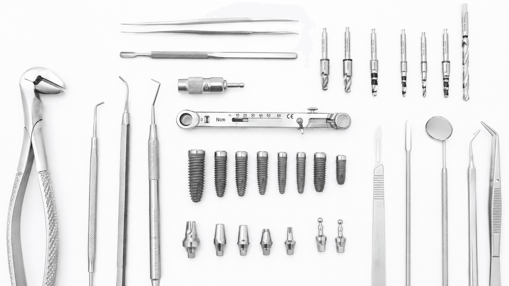
- Primary stability: Insertion torque of 25–45 Ncm is the clinical benchmark. Below 25 Ncm in poor bone quality, immediate loading is contraindicated.
Osseointegration in Periodontics
Periodontics and implantology share the same biological ground: both fields depend on the health of the tissue — periodontal ligament for natural teeth, bone-to-implant contact for implants — that anchors a tooth-like structure under load. A periodontist evaluating a patient for implant therapy is assessing whether the remaining bone and soft tissue can support osseointegration in the first place — active periodontal disease, untreated, is one of the most common reasons osseointegration fails or peri-implantitis develops later.
Clinical Takeaway
For the dentist transitioning into implantology, osseointegration is not a passive process you wait for — it is an outcome you engineer through patient selection, site preparation, and implant system choice. Understanding the biology is what separates a confident implantologist from one who places and hopes.